PNEUMATA
Interactive 3D anatomical model — 37 organ nodes mapped to hardware analogs, 24 vertebral disc markers, bidirectional spine highlighting, four view modes.
About
The human body and a computer are not metaphorically similar — they are architecturally isomorphic. The heart is a PSU. The spinal cord is a PCIe bus. The kidneys are a virtual memory manager. The immune system is an IDS with edge nodes, a SIEM, and a definition update engine.
Pneumata makes that argument visual and interactive. Every organ node corresponds to a specific hardware component. Every spinal disc corresponds to a bus arbitration layer. The relationship is functional, not decorative — the same engineering problem solved twice, in different substrates, separated by 500 million years.
Node Placement
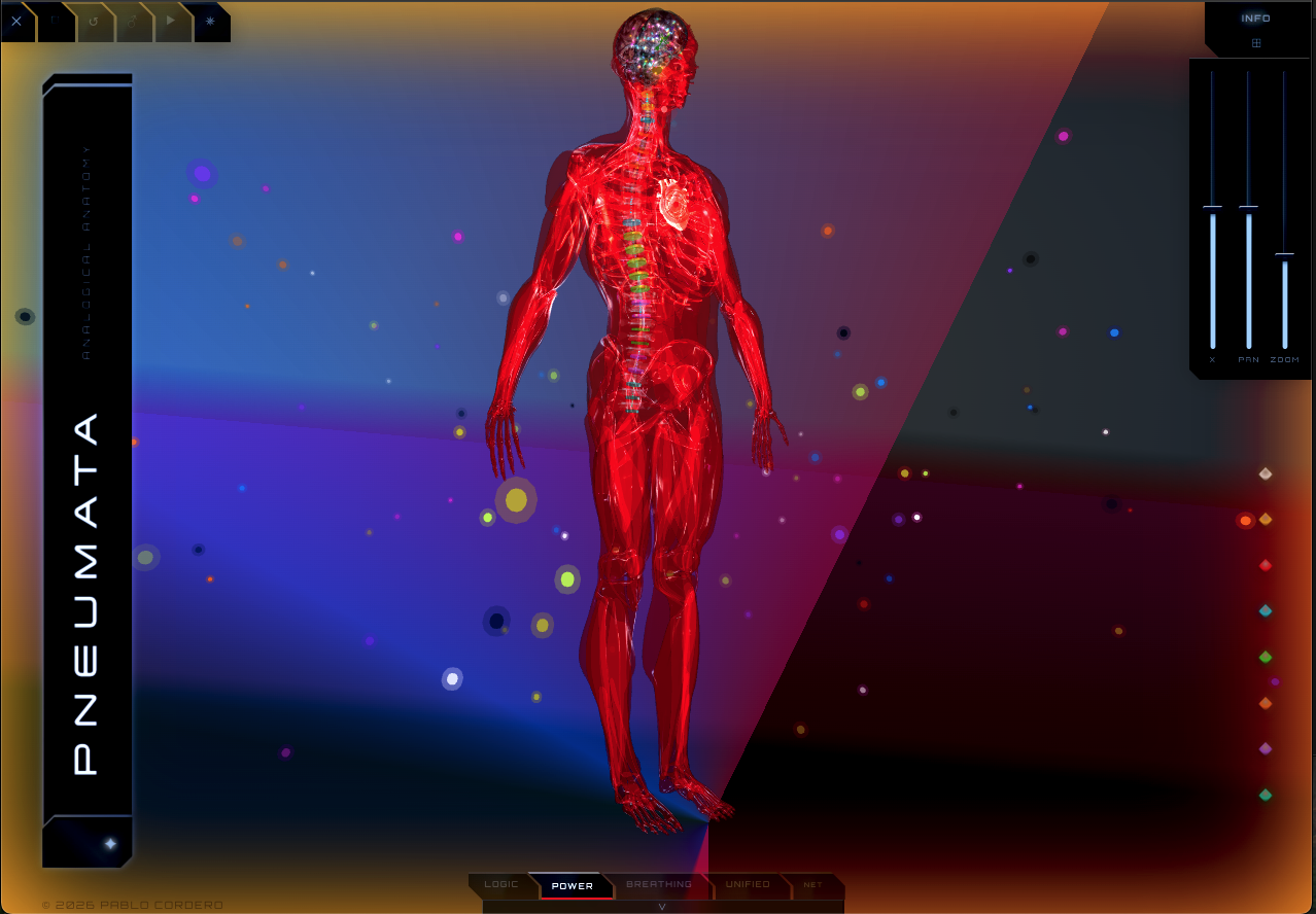
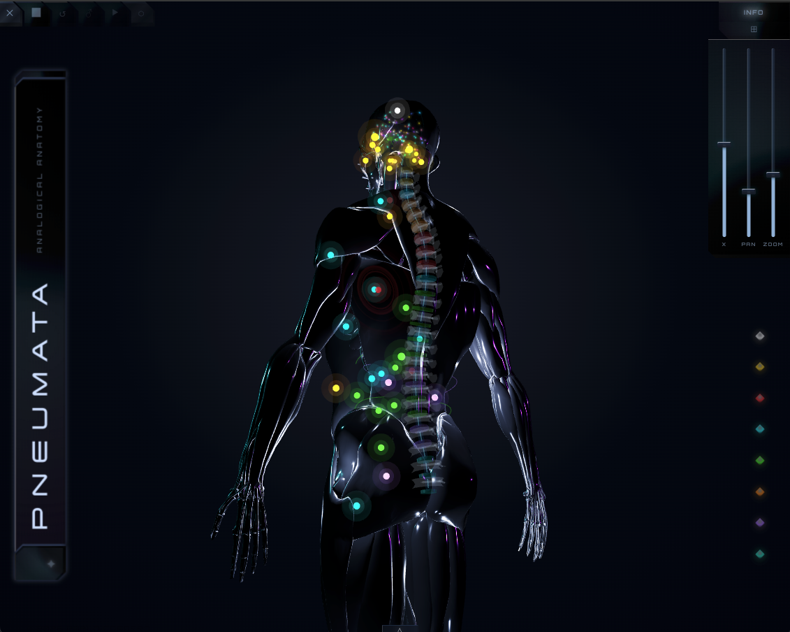
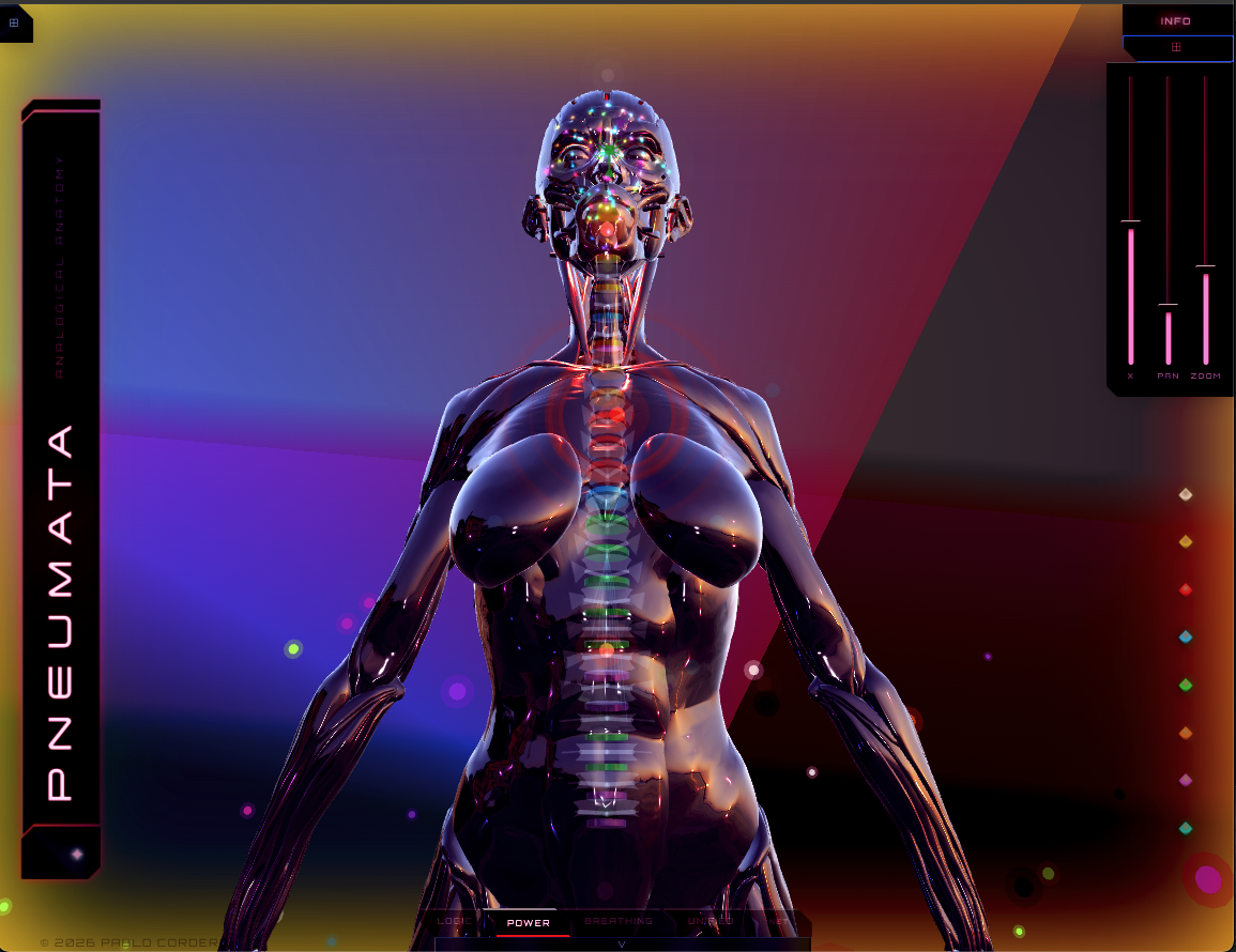
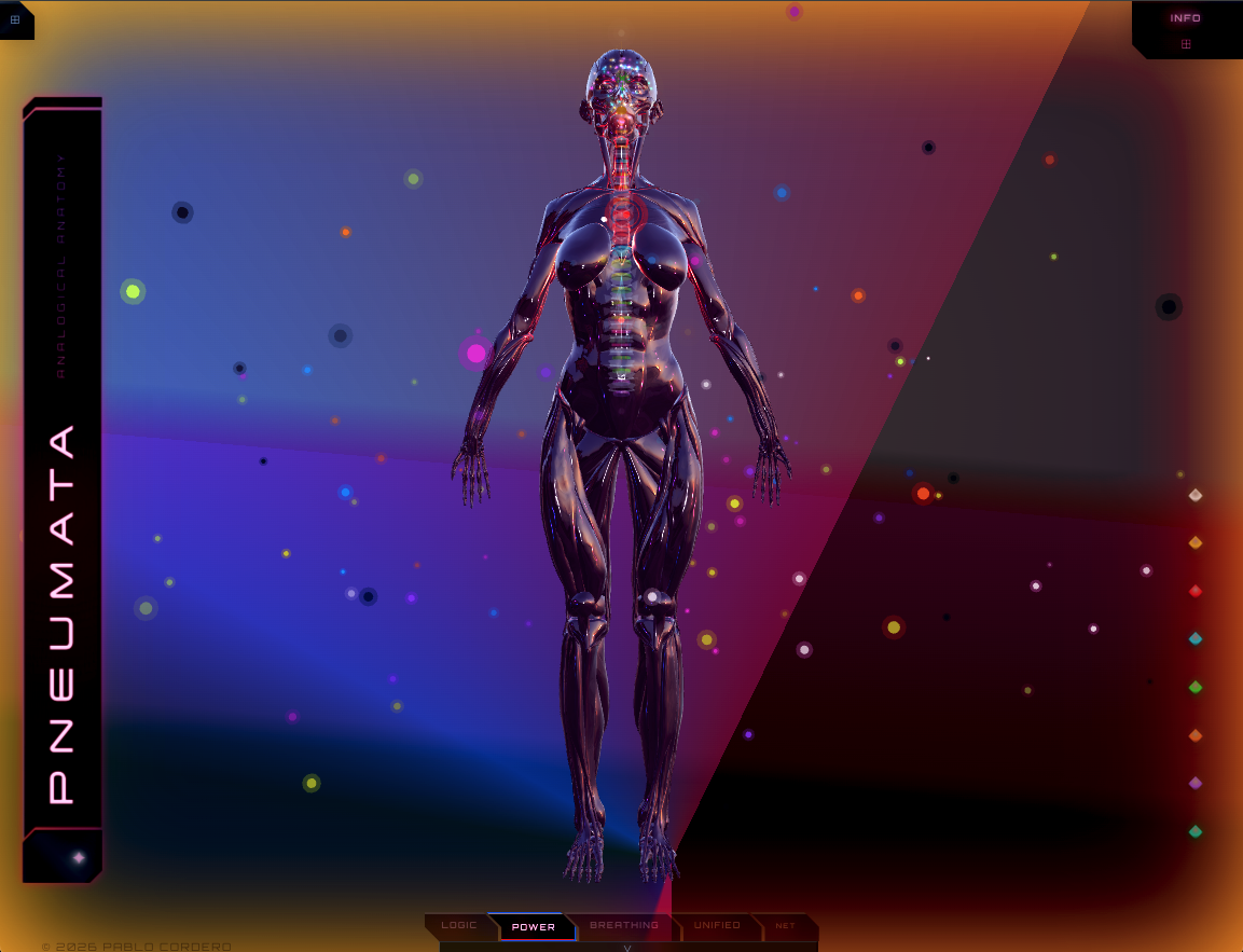
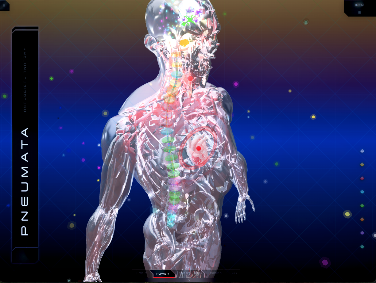
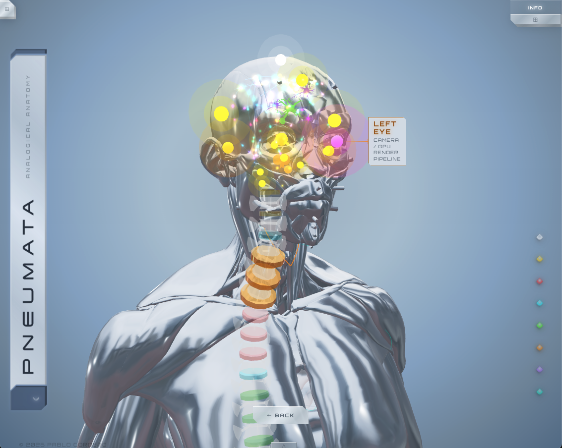
37 organ nodes are positioned in 3D space using vertex-sampled coordinates extracted directly from the GLB anatomical mesh at runtime — no hardcoded positions. Each belongs to one of 8 color-coded categories: Logic, Power, Thermal, Digestive, Sensory, Renal, Immune, Spirit.
The spinal column is auto-traced using a y-band bucketing algorithm across the mesh's vertex data. 24 vertebral disc markers (C2 through S2) are distributed along that traced geometry and color-coded by the organ system innervated at each spinal level — derived from actual anatomical innervation data.
Bidirectional Signaling
Hover an organ node — its spinal bus lanes illuminate. Hover a
disc — its innervated organs respond in kind. Signal propagates
both directions simultaneously, the way nerve conduction actually
does. This is implemented through a single
hoveredCategory state consumed by both
OrganNode and SpinalCord in parallel —
no prop drilling, no global store.
View Modes
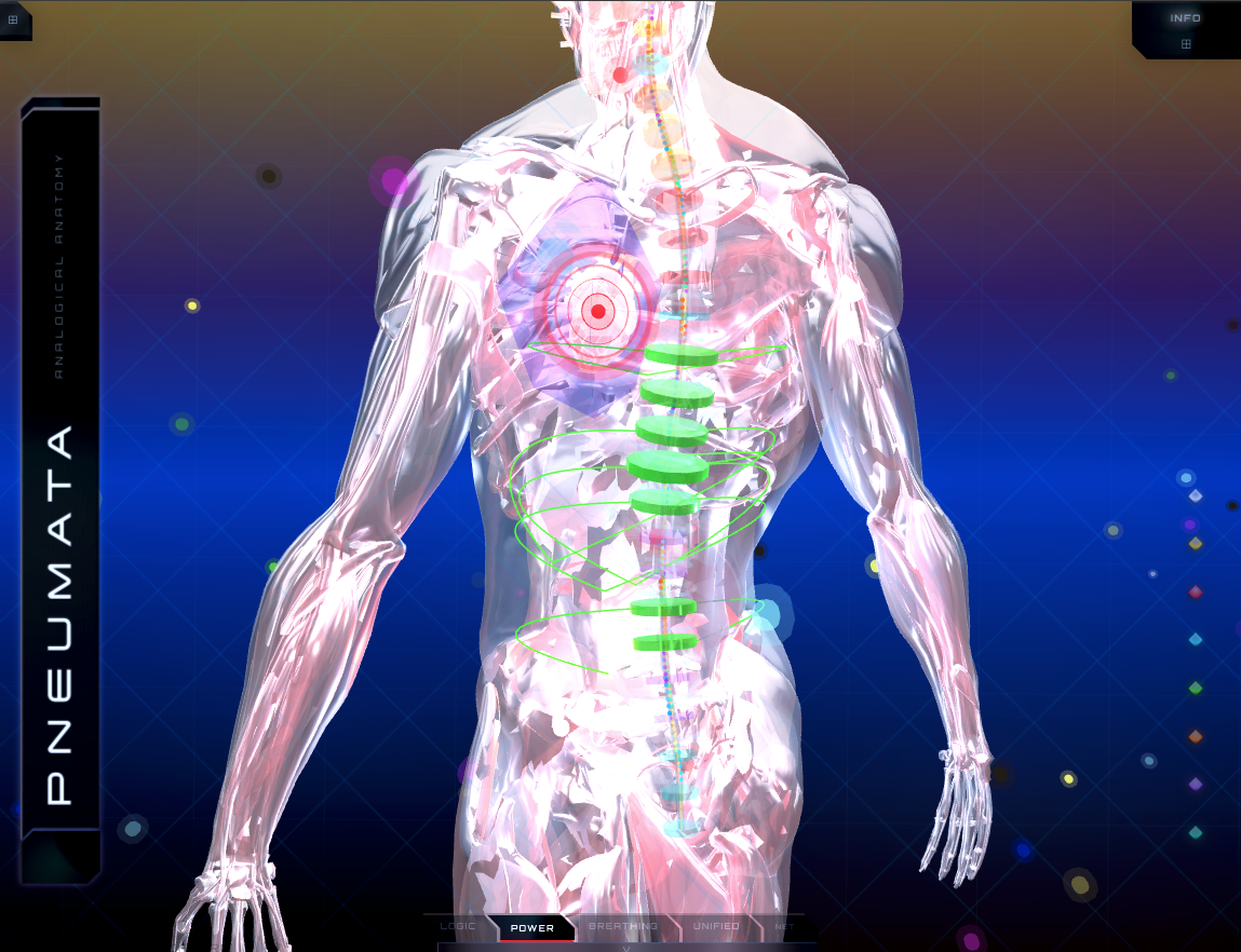
Four modes, each activating a different system. Logic — the neural network: processing, memory, signal. Power — two energy orbs circuit the full body while the heart node radiates rings each clock cycle. Breathing — the mesh shifts from blue to cyan at 0.25Hz, lung nodes flashing at oxygenation. Unified — all systems simultaneously.
The User
One node breaks the hardware pattern on purpose. Pneuma, the consciousness node, isn't mapped to a component — its hardware analog is labeled "The User." A computer runs whether or not anyone is watching; a body doesn't appear to. Federico Faggin, who helped invent the microprocessor at Intel, spent his later career arguing consciousness precedes computation rather than emerging from it. Pneumata's philosophy sides with him.
Tech Stack
My Role
Solo — 3D scene architecture, organ data system, spinal auto-trace algorithm, bidirectional highlight logic, view mode animations, modal system, and all UI.
Project Type
Interactive 3D Visualization / Philosophy
Timeline
March – April 2026
System Architecture
37 Organ Nodes
Every major organ mapped to a hardware analog across 8 categories. Positions sampled from GLB mesh vertex data at runtime — no hardcoded coordinates, no guesswork.
Auto-Traced Spine
Vertebral column geometry extracted from the GLB mesh at runtime via vertex filtering and y-band bucketing. 24 disc markers placed along the real anatomical curve, C2 through S2.
Bidirectional Highlighting
Hover an organ — its spinal bus lanes light up. Hover a disc — its innervated organs glow back. Category color propagates in both directions, mirroring the bidirectionality of nerve conduction.
Traveling Circulation Orbs
Two energy nodes circuit the full body on closed loops in Power mode. The heart radiates expanding rings on each pass — a PSU distributing charge across the network on every clock cycle.
Breathing Mode
The body mesh cycles blue (deoxygenated, inhale) to cyan (oxygenated, peak) at 0.25Hz. Lung nodes flash at the moment of oxygenation — the pulmonary system rendered as a live signal.
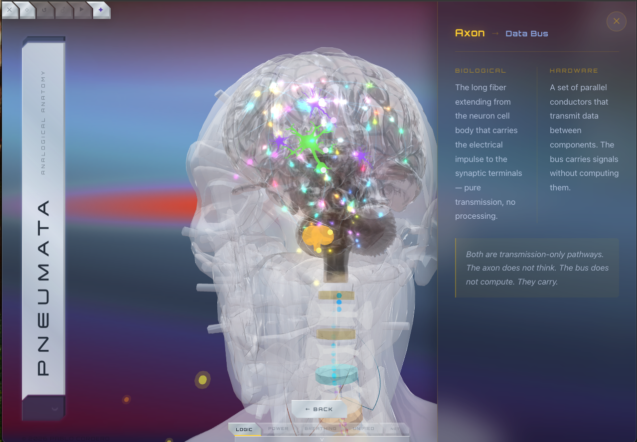
Organ + Disc Modals
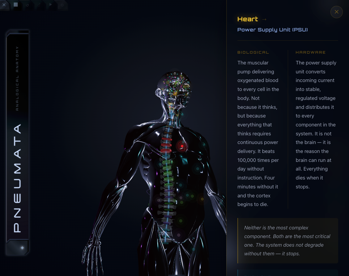
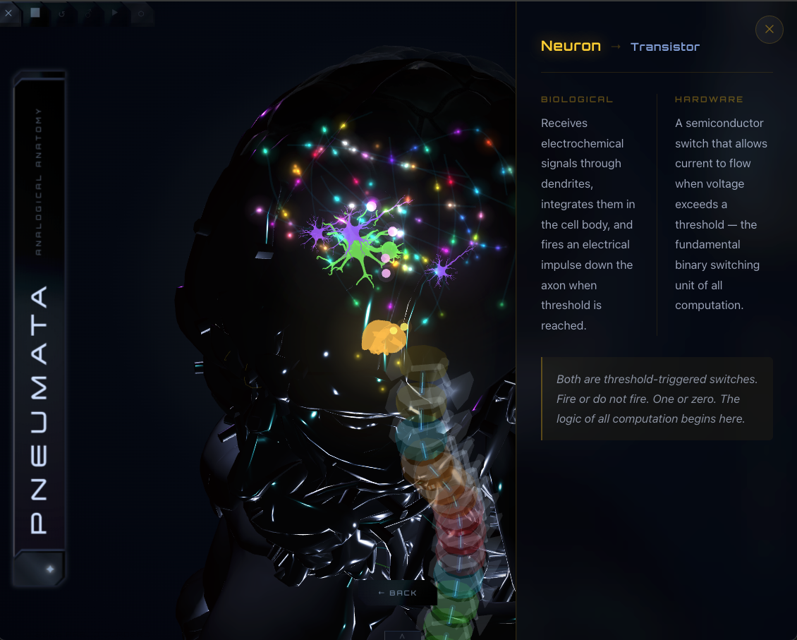
Clicking any node opens a panel with biological function, hardware analog, synthesis, and spinal bus lane assignment with PCIe lane analogy. Disc clicks surface level, innervation target, and arbitration layer description.
NET Overlay
Toggle the full peripheral nervous system: spinal nerve roots mapped from each disc to its innervated organs as curved Bézier paths, plus white matter tracts between brain regions rendered as CatmullRom curves.
Brain Zoom
Clicking any cranial node transitions the camera into a dedicated brain view, repositioning all brain-region nodes to a zoomed cluster. Brain tracts (corpus callosum, hypothalamic-pituitary axis, thalamocortical) render as distinct white matter paths.